Medical imaging plays a critical role in diagnosing, monitoring, and treating various medical conditions. From broken bones to complex neurological disorders, imaging technologies allow doctors to see inside the body without invasive procedures. In this post, we’ll explore the most common types of medical imaging, how they work, their applications, and what to expect during each procedure.
1. X-Ray (Radiography)
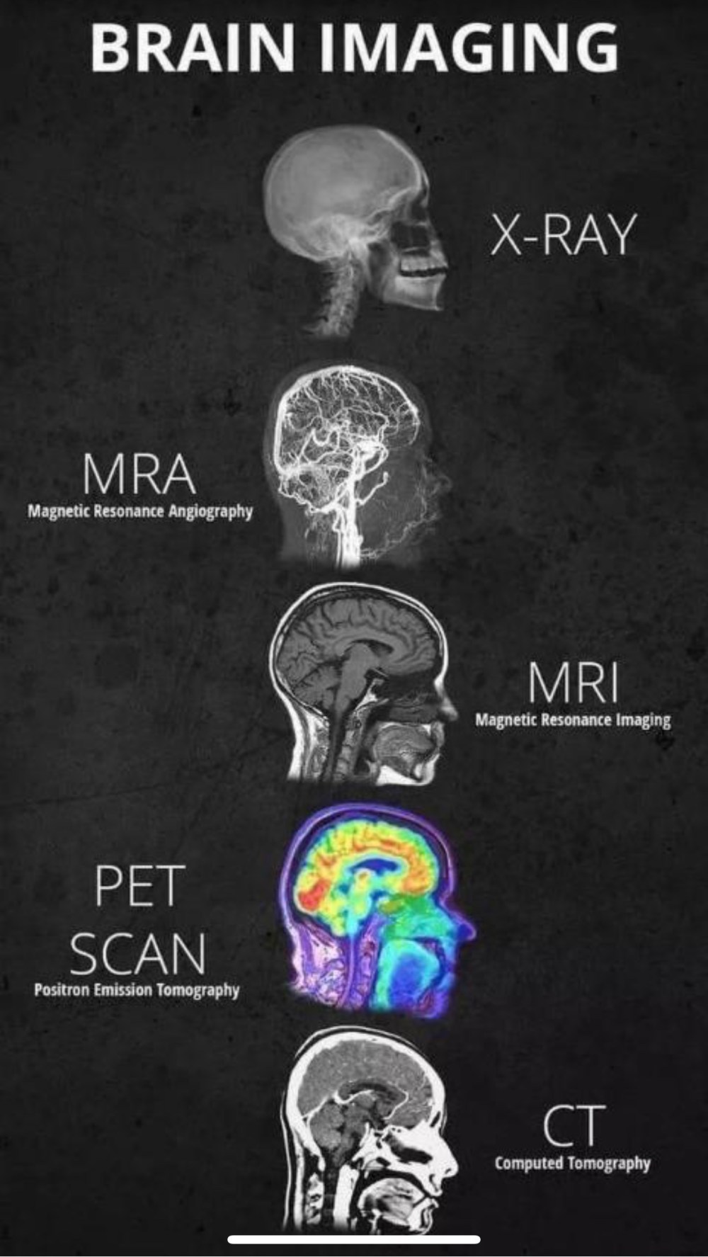
How It Works: X-rays use a small amount of radiation to create images of structures inside the body. Dense materials, like bones, absorb more radiation and appear white on the image, while soft tissues appear darker.
Common Uses:
- Diagnosing fractures and dislocations
- Detecting lung conditions (e.g., pneumonia, tuberculosis)
- Dental imaging for cavities and alignment
What to Expect:
- The procedure is quick and painless.
- You may need to hold a specific position or wear a lead apron to protect other areas from radiation.
2. Computed Tomography (CT or CAT Scan)
How It Works: CT scans combine X-rays with computer processing to create detailed cross-sectional images of the body. They provide more detail than standard X-rays.
Common Uses:
- Detecting tumors or cancers
- Diagnosing internal injuries or bleeding
- Evaluating complex bone fractures
- Guiding biopsies or surgeries
What to Expect:
- You’ll lie on a table that slides into a large, doughnut-shaped machine.
- Some scans require contrast dye for clearer images, administered orally or intravenously.
- The procedure usually takes 10–30 minutes.
3. Magnetic Resonance Imaging (MRI)
How It Works: MRIs use powerful magnets and radio waves to create detailed images of organs and soft tissues. Unlike X-rays and CT scans, MRIs don’t use radiation.
Common Uses:
- Diagnosing brain and spinal cord conditions
- Identifying ligament or tendon injuries
- Monitoring organ damage or diseases
- Detecting tumors in soft tissues
What to Expect:
- You’ll lie inside a large, tube-like machine.
- The machine makes loud noises, so earplugs or headphones may be provided.
- The procedure is non-invasive but can take 30–60 minutes.
4. Ultrasound (Sonography)
How It Works: Ultrasound uses high-frequency sound waves to create real-time images of organs, tissues, and blood flow. It’s a radiation-free method.
Common Uses:
- Monitoring pregnancy and fetal development
- Evaluating blood flow in veins and arteries
- Diagnosing abdominal or pelvic conditions
- Guiding minimally invasive procedures
What to Expect:
- A handheld device (transducer) is moved over the skin, often with a gel applied to enhance contact.
- It’s painless and typically takes 20–40 minutes.
5. Positron Emission Tomography (PET Scan)
How It Works: PET scans use a small amount of radioactive tracer to highlight areas of high metabolic activity, often indicative of disease.
Common Uses:
- Detecting cancer and monitoring its spread
- Evaluating brain disorders (e.g., Alzheimer’s, epilepsy)
- Assessing heart function and blood flow
What to Expect:
- A radioactive tracer is injected or swallowed before the scan.
- You’ll lie on a table that moves through a scanner.
- The procedure takes about 30–60 minutes.
6. Mammography
How It Works: Mammograms use low-dose X-rays to create images of breast tissue, helping detect early signs of breast cancer.
Common Uses:
- Routine breast cancer screening
- Diagnosing breast lumps or changes
What to Expect:
- The breast is positioned on a flat surface and compressed briefly to capture clear images.
- It may cause slight discomfort, but the procedure is quick.
7. Fluoroscopy
How It Works: Fluoroscopy provides real-time X-ray images, often enhanced with contrast dye, to observe internal motion.
Common Uses:
- Guiding catheter insertions (e.g., for heart or vascular procedures)
- Evaluating digestive tract issues (e.g., swallowing difficulties)
- Performing joint injections or spinal treatments
What to Expect:
- The procedure may involve swallowing a contrast dye or having it injected.
- It’s generally painless, though some tests may feel invasive.
8. Nuclear Medicine Imaging
How It Works: Nuclear medicine involves injecting or ingesting a small amount of radioactive material, which is then detected by special cameras to create images of internal organs.
Common Uses:
- Assessing thyroid function
- Detecting bone abnormalities or fractures
- Evaluating kidney or heart function
What to Expect:
- You’ll receive a radioactive tracer and then wait for it to circulate.
- The scan is painless and can take 30 minutes to several hours, depending on the test.
9. Dual-Energy X-Ray Absorptiometry (DEXA or Bone Density Scan)
How It Works: DEXA uses low-dose X-rays to measure bone density, helping diagnose osteoporosis.
Common Uses:
- Assessing bone strength and fracture risk
- Monitoring bone health in people with osteoporosis
What to Expect:
- You’ll lie on a table while the scanner passes over your body.
- It’s quick, painless, and typically takes 10–20 minutes.
10. Echocardiography (Heart Ultrasound)
How It Works: This specialized ultrasound creates images of the heart, allowing doctors to assess its structure and function.
Common Uses:
- Diagnosing heart valve issues
- Assessing heart wall motion
- Evaluating heart defects
What to Expect:
- A transducer is placed on your chest or inserted into your esophagus (for transesophageal echocardiograms).
- It’s painless unless an invasive approach is required.
Choosing the Right Imaging Test
The type of imaging used depends on the condition being evaluated. Your doctor will choose the most appropriate test based on your symptoms, medical history, and the area of concern.
Final Thoughts
Medical imaging has revolutionized healthcare, enabling earlier and more accurate diagnoses. While the machines and procedures may seem intimidating, most are non-invasive and straightforward. Remember, these technologies are here to provide valuable insights and guide your treatment.
Have you had any imaging tests done? Share your experiences or questions in the comments below!
Note: Always consult a healthcare professional for guidance on medical imaging and your health concerns. This post is for informational purposes only.
